Cryo-Electron Tomography of Archaeal Biofilm Architectures: Nanoscale Insights into Extremophile Adaptation and Bioremediation Strategies
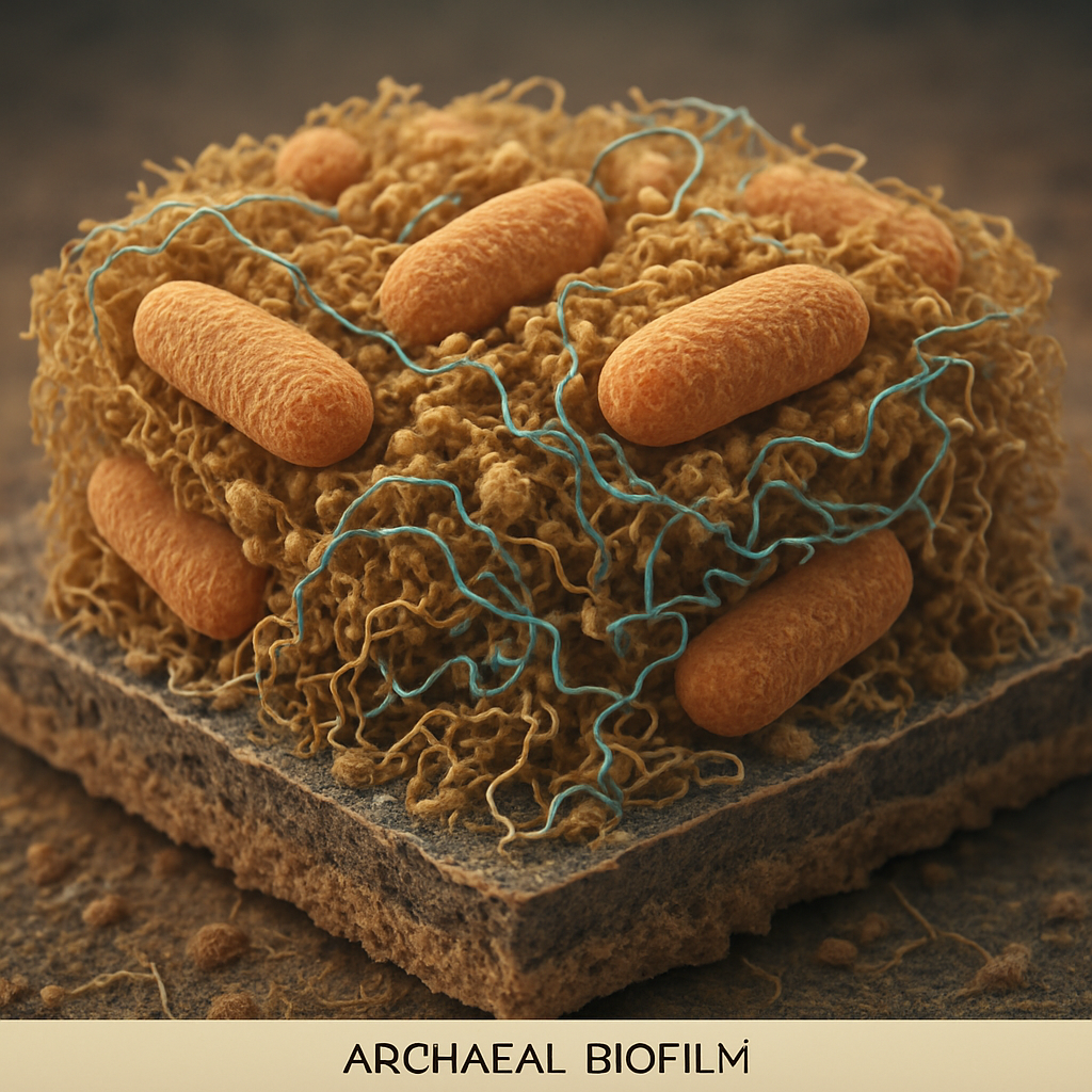
Archaeal biofilms, particularly those formed by extremophiles, represent fascinating and resilient microbial communities capable of thriving in environments hostile to most life forms. These complex architectures are crucial for archaeal survival, facilitating nutrient acquisition, protection from stressors, and intercellular communication. Understanding the nanoscale organization of these biofilms is paramount for deciphering the adaptive strategies of extremophiles and for harnessing their unique capabilities in bioremediation and biotechnology. Cryo-electron tomography (cryo-ET) has emerged as a powerful technique, enabling the visualization of these intricate structures in their near-native, hydrated state, providing unprecedented insights into their formation, composition, and function.
Nanoscale Architecture and Extracellular Matrix of Archaeal Biofilms
Cryo-ET studies are beginning to unveil the sophisticated three-dimensional organization of archaeal biofilms. These investigations reveal densely packed cellular arrangements often encased in an extensive extracellular matrix (ECM). The ECM, a hallmark of biofilm communities, is a complex meshwork of extracellular polymeric substances (EPS), including proteins, polysaccharides, lipids, and potentially extracellular DNA (eDNA). For instance, studies on Methanospirillum hungatei have used cryo-ET to reveal the hierarchical organization of its proteinaceous sheath, which is composed of amyloid-like proteins forming stacked rings of β-strand arches, providing structural integrity to the cells (Wang et al., 2023). While not strictly biofilms, these sheaths offer insights into archaeal extracellular structures. The composition and architecture of the ECM in extremophilic archaeal biofilms are hypothesized to be specifically adapted to the extreme conditions they inhabit, such as high temperatures, extreme pH, or high salinity. For example, halophilic archaea like Halobacterium salinarum have been studied using cryo-EM techniques, highlighting strategies to adapt cell envelopes to high salt concentrations, which directly impacts biofilm formation and stability (Bollschweiler et al., 2017). The intricate network of the ECM likely plays a critical role in protecting cells from desiccation, UV radiation, toxic compounds, and phagocytosis, while also contributing to the biofilm's mechanical stability and adhesion to surfaces.
Cellular Interactions and Appendages in Extremophile Adaptation
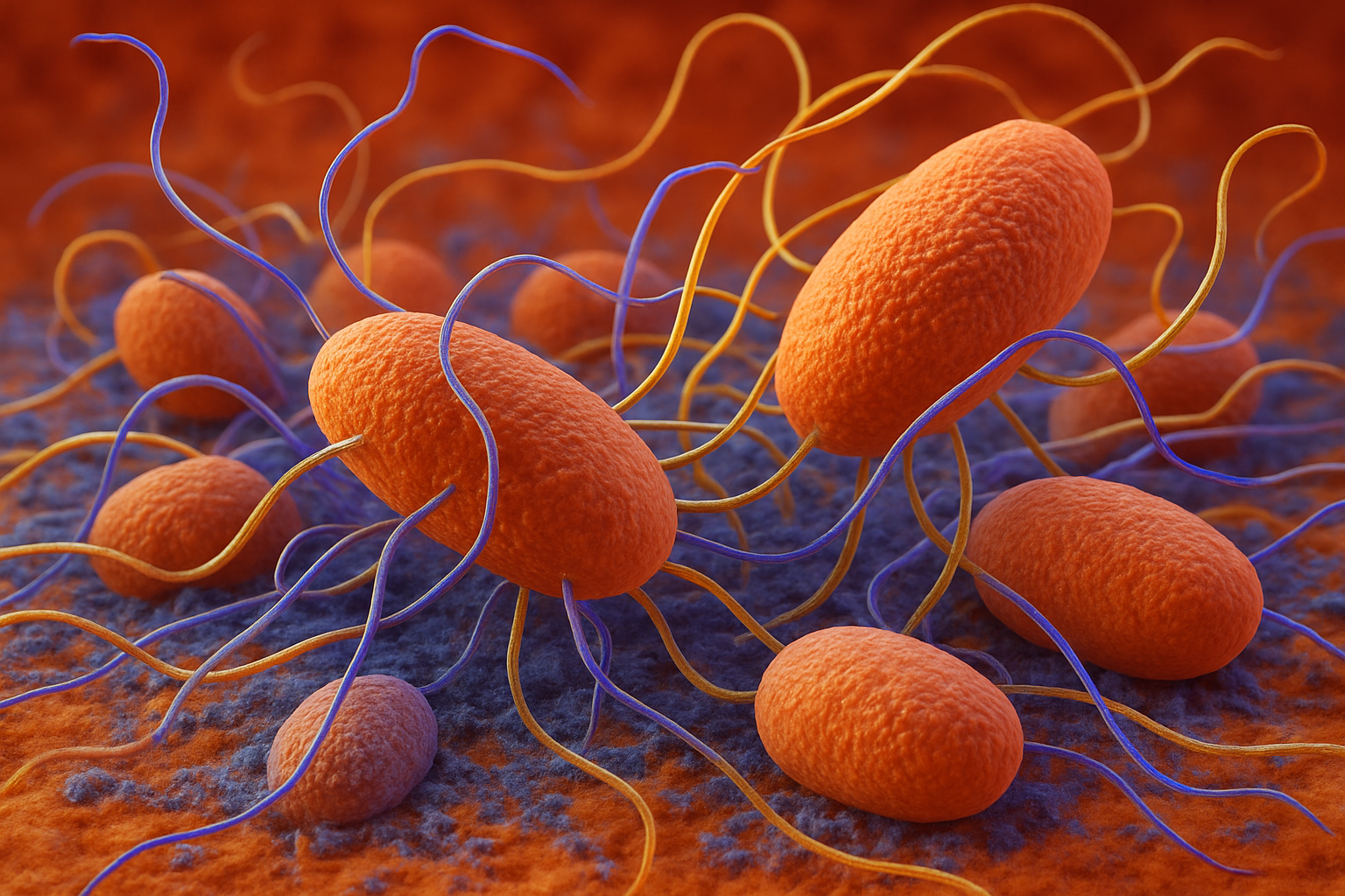
Within archaeal biofilms, cells exhibit complex interactions mediated by various surface structures and appendages, crucial for biofilm development and function in extreme environments. Cryo-ET can visualize these structures, such as pili, flagella (archaella), and other cell surface proteins, in situ. For example, retractable Type IV pili have been shown to mediate twitching motility in Sulfolobus acidocaldarius, a hyperthermophilic archaeon, enabling surface colonization under extreme conditions (Charles-Orszag et al., 2024). While not all archaea possess archaella for motility, other surface appendages are likely involved in cell-cell adhesion and biofilm maturation. Comparative genomics of archaea from acid mine drainage (AMD) biofilms, such as Ferroplasma spp. and related Thermoplasmatales, has revealed genes for pili production, and cryo-ET has corroborated these predictions, suggesting their role in biofilm structure and intercellular connections within these low-pH, metal-rich environments (Yelton et al., 2013). Understanding the spatial arrangement and molecular identity of these appendages will shed light on how archaeal cells communicate, exchange genetic material, and coordinate behavior within the biofilm, all critical for survival and adaptation in fluctuating extreme environments.

Bioremediation Potential and Biofilm Engineering Insights
The unique metabolic capabilities of extremophilic archaea, often amplified within the protective biofilm niche, make them promising candidates for bioremediation of contaminated environments and for various biotechnological applications. Archaeal biofilms can degrade recalcitrant pollutants, immobilize heavy metals, or produce novel biomolecules. For example, studies on hot spring biofilms highlight the role of diverse microbial taxa, including archaea, in the degradation of complex carbohydrates like starch, cellulose, and hemicellulose at elevated temperatures (Liew et al., 2024). The nanoscale insights gained from cryo-ET into biofilm architecture and ECM composition can inform strategies for engineering robust and efficient archaeal biofilms for targeted bioremediation. For example, understanding how the ECM sequesters metals or facilitates enzymatic activity could lead to the design of biofilm-based bioreactors. Furthermore, insights into the microbial diversity and functional adaptations within these biofilms, such as those found in saline-alkali soils and salt lakes (Ding et al., 2025), can reveal novel metabolic pathways and enzymes. The study by Chia et al. (2024) emphasizes the role of extremophiles, including halophilic archaea, in degrading emerging pollutants, suggesting that understanding their biofilm structures could enhance these bioremediation strategies.
Conclusion
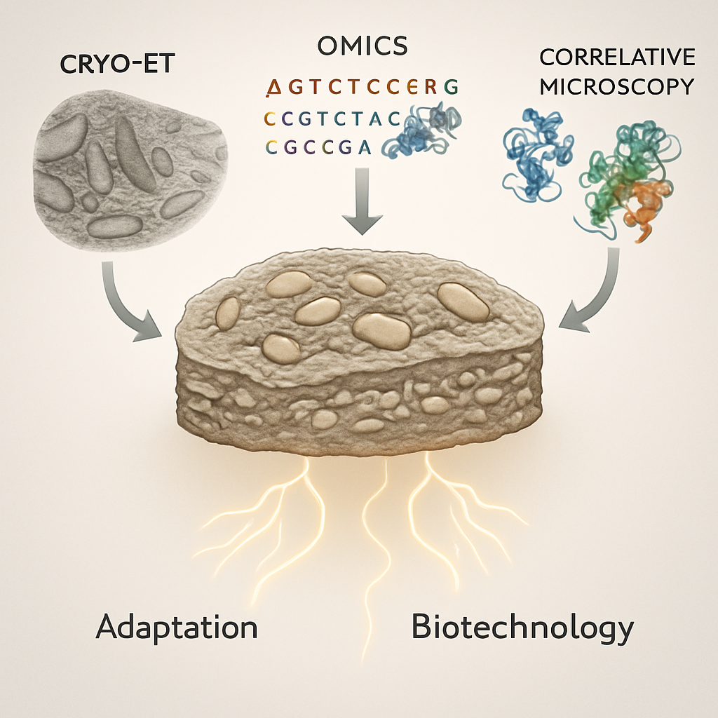
Cryo-electron tomography is revolutionizing our understanding of archaeal biofilm architectures, providing a crucial window into the nanoscale adaptations of extremophiles. Future cryo-ET studies, integrated with advanced omics techniques (genomics, transcriptomics, proteomics, metabolomics) and correlative light and electron microscopy (CLEM), will undoubtedly uncover further details about the molecular composition of the ECM, the dynamics of biofilm formation, intercellular signaling pathways, and the spatial organization of metabolic processes within these resilient communities. This deeper understanding will not only illuminate fundamental aspects of archaeal biology and microbial ecology in extreme environments but also catalyze the development of innovative bioremediation strategies. Harnessing the power of these natural extremophilic consortia, potentially through engineered biofilms with optimized architectures and functionalities, holds immense promise for addressing pressing environmental challenges and advancing biotechnology. The exploration of diverse extremophilic archaea and their biofilms using cryo-ET will continue to reveal nature's ingenuity in adapting life to the most inhospitable corners of our planet.
References
- Bollschweiler, D., Schaffer, M., Lawrence, C. M., & Engelhardt, H. (2017). Cryo-electron microscopy of an extremely halophilic microbe: technical aspects. Extremophiles, 21(2), 425-434. https://doi.org/10.1007/s00792-016-0912-0
- Charles-Orszag, A., Wolferen, M., Lord, S. J., Albers, S.-V., & Mullins, R. D. (2024). Adhesion pilus retraction powers twitching motility in the thermoacidophilic crenarchaeon Sulfolobus acidocaldarius. Nature Communications, 15(1), 4908. https://doi.org/10.1038/s41467-024-49101-7
- Chia, X. K., Hadibarata, T., Jusoh, M. N. H., Sutiknowati, L. I., Tan, I. S., & Foo, H. C. Y. (2024). Role of Extremophiles in Biodegradation of Emerging Pollutants. Topics in Catalysis. https://doi.org/10.1007/s11244-024-01919-7
- Ding, Y., Ke, J., Hong, T., Zhang, A., Wu, X., Jiang, X., Shao, S., Gong, M., Zhao, S., Shen, L., & Chen, S. (2025). Microbial diversity and ecological roles of halophilic microorganisms in Dingbian (Shaanxi, China) saline-alkali soils and salt lakes. BMC Microbiology, 25(1), 133. https://doi.org/10.1186/s12866-025-03997-3
- Liew, K. J., Shahar, S., Shamsir, M. S., Shaharuddin, N. B., Liang, C. H., Chan, K.-G., Pointing, S. B., Sani, R. K., & Goh, K. M. (2024). Integrating multi-platform assembly to recover MAGs from hot spring biofilms: insights into microbial diversity, biofilm formation, and carbohydrate degradation. Environmental Microbiome, 19(1), 35. https://doi.org/10.1186/s40793-024-00572-7
- Wang, H., Zhang, J., Toso, D., Liao, S., Sedighian, F., Gunsalus, R., & Zhou, Z. H. (2023). Hierarchical organization and assembly of the archaeal cell sheath from an amyloid-like protein. Nature Communications, 14(1), 6790. https://doi.org/10.1038/s41467-023-42368-2
- Yelton, A. P., Comolli, L. R., Justice, N. B., Castelle, C., Denef, V. J., Thomas, B. C., & Banfield, J. F. (2013). Comparative genomics in acid mine drainage biofilm communities reveals metabolic and structural differentiation of co-occurring archaea. BMC Genomics, 14, 485. https://doi.org/10.1186/1471-2164-14-485